
Western researchers bring X-ray technology to developing countries
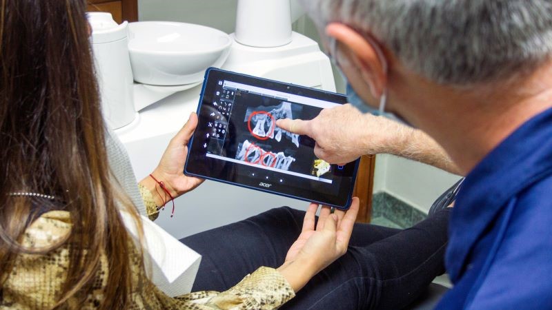
Roentgen discovered X-rays well over a century ago, yet nearly two-thirds of the world’s population has no access to diagnostic medical imaging, according to the World Health Organization. A startling statistic, says David Holdsworth, professor of surgery and medical biophysics in the Schulich School of Medicine & Dentistry, and one that needs to be rectified. To do just that, Bone and Joint Institute (BJI) member Holdsworth and a team of African and Canadian medical imaging experts are in the process of developing a cost-effective, portable digital X-ray system. It will help clinicians in low- and middle-income countries to provide better medical care and prevent broken bones from growing into long-term health issues.
“Road accidents are on the rise in low- and middle-income countries, and that means muscle and bone injuries are on the rise, too. Accidents account for over 90 percent of serious injuries,” says Holdsworth. “If someone breaks a bone but can’t get an X-ray at their rural clinic, that can lead to the fracture not setting correctly and subsequent long-term disability,” says Holdsworth.
The scarcity of X-ray technology in developing countries stems from many reasons: from civil unrest, to restricted resources, or lack of reliable power. In addition, complex imaging devices that need to be operated by specialists aren’t a good fit for rural and remote areas. That’s why developing an X-ray system that is easy to build, maintain, and operate, is key in increasing equitable and wide-spread access to X-ray technology, says Holdsworth: “The solution is not to send obsolete equipment that we don't want anymore. It's not even collecting money and sending new equipment in boxes. It's working to develop systems that can be made and distributed and maintained locally.”
Holdsworth’s "bootstrap” imaging system has the potential to produce high quality digital X-ray images with minimal equipment: a portable X-ray tube, a phosphor screen, and a digital camera. State-of-the-art digital cameras are equipped with extremely sensitive image sensors that can transform even the smallest amount of light – such as the glow coming off a phosphor screen – into high quality images. Holdsworth’s plan involves leveraging those advances in camera technology to capture digital X-ray images that give traditional imaging machines a run for their money.
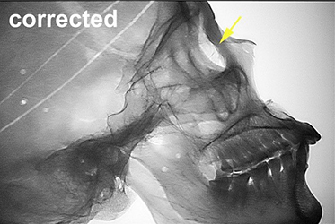
To verify his proposed X-ray system, Holdsworth tested his theory with a prototype system and manged to produce high-quality X-ray images of a human skull.
Holdsworth also just got news that he secured a competitive (<5 percent success rate) NSERC Discovery Horizons grant for $480,000.
Both the grant and the idea for the X-ray system were accelerated by a Bone and Joint Institute catalyst grant in 2020, he says. The grant supported the work of Holdsworth and biological anthropology professor and BJI member Jay Stock in building a portable CT and X-ray scanner to image human bones found at archeological sites.
“You can’t just take artifacts and put them in your suitcase and scan them at home. They are protected. But there are no scanners on site. In talking about this problem, I said something like, ‘Well, you could just use a camera and phosphor and you could probably get a scan.’ And through the BJI grant, we were able to set up a system to acquire X-ray images of bones. And they looked really good.”
Having already conducted research that proved his theory helped tremendously in securing additional funding for related projects, Holdsworth says: “We already had data from our Bone and Joint catalyst grant demonstrating the proof of principle. And we had images. I mean, you don’t have things like that just lying around in the timescale that you have to put one of these grants together.”
Looking towards the future, he hopes that his portable X-ray system will find many uses that serve people and places around the world, while raising Western’s profile as a leader in health and equity research: “I think that Western has the potential to become a centre of excellence for research that fosters health and equity in remote, rural and low-income countries. It’s a huge area of growth and there are so many opportunities.”